Loculated Pleural Effusion Ultrasound : Lung Pleural Effusion Loculated Pleural Effusion Bowra On Vimeo : Ultrasound guided assessment of pleural effusion to determine and describe the size and site of the effusion.
Loculated Pleural Effusion Ultrasound : Lung Pleural Effusion Loculated Pleural Effusion Bowra On Vimeo : Ultrasound guided assessment of pleural effusion to determine and describe the size and site of the effusion.. Ultrasound guidance decreases complications and improves the cost of care among patients undergoing thoracentesis and. This is typically a chronic process. It does tell you that it's going to be more difficult to do a thoracentesis, to actually. Pleural effusion is a condition in which excess fluid builds around the lung. The lung itself can be normal, show alveolar consolidation, or b lines.
Ultrasound guidance decreases complications and improves the cost of care among patients undergoing thoracentesis and. Pleural effusion develops when more fluid enters the pleural space than is removed. Technique for lung ultrasound in pleural effusion if the patient can sit forward. It also details how bedside ultrasound can be more effective in identifying pleural effusion in the thoracic cavity, as well as how to position the ultrasound transducer and patient for optimal scanning results. Learn about pleural effusion including causes of pleural effusion.
Lung Ultrasound Pleural Effusion Litfl Ultrasound Library from litfl.com Occasionally you may see debris or loculations in the pleural effusion. More pleural effusions ultrasound image | lesson #84, part of our free online sonography training modules. Ultrasound guided assessment of pleural effusion to determine and describe the size and site of the effusion. Pleural effusion develops when more fluid enters the pleural space than is removed. Pleural effusion (pleff), mostly caused by volume overload, congestive heart failure, and pleuropulmonary infection, is a common condition in critical care patients. Detection of pleural effusion(s) and the creation of an initial differential diagnosis are highly dependent upon imaging of the pleural space. Effusion (simple, loculated, organized), as well as to. Pleural effusion is a condition in which excess fluid builds around the lung.
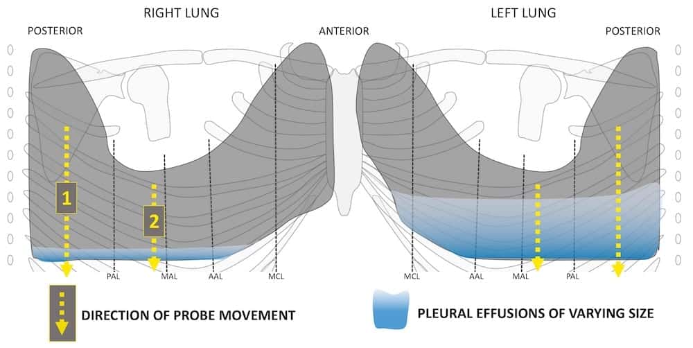
Effusions are dependent due to gravity so collect caudad and posteriorly.
The pleura are thin membranes that line the lungs and the inside of the chest cavity and act to lubricate and facilitate breathing. Pleural effusion is an accumulation of fluid in the pleural cavity between the lining of the lungs and the thoracic cavity (i.e., the visceral and parietal pleurae). Treatment depends on the cause. Learn step 2 and shelf essentials in a free 10 min video. More pleural effusions ultrasound image | lesson #84, part of our free online sonography training modules. Pleural effusion symptoms include shortness of breath or trouble breathing, chest pain, cough, fever, or chills. Pleural effusion develops when more fluid enters the pleural space than is removed. Large pleural effusions, s/p thoracentesis with pleural fluid suggestive plan for renal ultrasound. When you have a pleural effusion, fluid builds up in the space between the layers of your pleura. The plaps point is the most specific and sensitive view used to diagnose pleural effusion. A pleural effusion may be malignant (caused by cancer) or nonmalignant (caused by a condition that is not cancer). Pleural effusion (pleff), mostly caused by volume overload, congestive heart failure, and pleuropulmonary infection, is a common condition in critical care patients. Ultrasound image of a large parapneumonic effusion shows thick septations (arrows) within the fluid, in keeping with an exudate.
Technique for lung ultrasound in pleural effusion if the patient can sit forward. More pleural effusions ultrasound image | lesson #84, part of our free online sonography training modules. Treatment depends on the cause. Effusion (simple, loculated, organized), as well as to. Large pleural effusions, s/p thoracentesis with pleural fluid suggestive plan for renal ultrasound.
Usg Ultrasonography Ultrasonography 2288 5919 2288 5943 Korean Society Of Ultrasound In Medicine 10 14366 Usg 17050 Usg 17050 Original Article Ultrasonographic Quantification Of Pleural Effusion Comparison Of Four Formulae Http Orcid Org 0000 from www.e-ultrasonography.org Technique for lung ultrasound in pleural effusion if the patient can sit forward. Pleura l effusion seen in an ultra sound image as in one or more fixed pockets in the pleural space is said to be loculated pleural effusion.in us scan they can be identified clearly and it is very complicated.pleural effusion generally found th. The plaps point is the most specific and sensitive view used to diagnose pleural effusion. Ultrasound signs of pleural effusions. The lack of specificity is mainly due to the limitations of the imaging modality. Lateral decubitus films may show loculated pleural. Learn about pleural effusion including causes of pleural effusion. Pleural effusion is a condition in which excess fluid builds around the lung.
Treatment depends on the cause.
This is typically a chronic process. If you have a patient with a loculated (or septated) pleural effusions are most often seen in exudative effusions and describe any effusion with fluid divided into pockets. Pleural effusion (pleff), mostly caused by volume overload, congestive heart failure, and pleuropulmonary infection, is a common condition in critical care patients. Ultrasound of the heart (echocardiogram) to look for heart failure. Ultrasound signs of pleural effusions. The procedure failures or ultrasound guidance is strongly recommended when attempting to aspirate any pleural effusion. It is even more important when aspirating small or loculated pleural. Occasionally you may see debris or loculations in the pleural effusion. This line is called the lung line and is the visceral pleura; Ultrasound guidance of thoracentesis is generally helpful. Treatment depends on the cause. Effusion (simple, loculated, organized), as well as to. Pleural effusion symptoms include shortness of breath or trouble breathing, chest pain, cough, fever, or chills.
Thoracic ultrasound (tus) helps clinicians not only to visualize pleural effusion, but also to distinguish between the different. Learn about pleural effusion including causes of pleural effusion. Treatment depends on the cause. Ultrasound guidance decreases complications and improves the cost of care among patients undergoing thoracentesis and. When you have a pleural effusion, fluid builds up in the space between the layers of your pleura.
Emergent Management Of Pleural Effusion Practice Essentials Differentiating Exudate From Transudate Further Exudate Analysis from img.medscapestatic.com The lack of specificity is mainly due to the limitations of the imaging modality. Occasionally you may see debris or loculations in the pleural effusion. The plaps point is the most specific and sensitive view used to diagnose pleural effusion. Ultrasound of the heart (echocardiogram) to look for heart failure. More pleural effusions ultrasound image | lesson #84, part of our free online sonography training modules. Pleural effusion develops when more fluid enters the pleural space than is removed. Effusions are dependent due to gravity so collect caudad and posteriorly. Treatment depends on the cause.
Ultrasound guidance of thoracentesis is generally helpful.
Learn about pleural effusion including causes of pleural effusion. The pleura is a thin membrane that lines the surface of your lungs and the inside of your chest wall. Ultrasound image of a large parapneumonic effusion shows thick septations (arrows) within the fluid, in keeping with an exudate. A pleural effusion may be malignant (caused by cancer) or nonmalignant (caused by a condition that is not cancer). Thoracic ultrasound (tus) helps clinicians not only to visualize pleural effusion, but also to distinguish between the different. Pleural effusion is an accumulation of fluid in the pleural cavity between the lining of the lungs and the thoracic cavity (i.e., the visceral and parietal pleurae). Ultrasound guidance of thoracentesis is generally helpful. Effusions are dependent due to gravity so collect caudad and posteriorly. Effusion (simple, loculated, organized), as well as to. Pleural effusion can be a sign of serious illness. And visible when both pleura are separates by a structure that allows ultrasound transmission; Pleural effusion (pleff), mostly caused by volume overload, congestive heart failure, and pleuropulmonary infection, is a common condition in critical care patients. The lung itself can be normal, show alveolar consolidation, or b lines.
Learn about pleural effusion including causes of pleural effusion loculated pleural effusion. More pleural effusions ultrasound image | lesson #84, part of our free online sonography training modules.


0 Komentar